
Fibrosis Models
Advanced fibrosis models that replicate key disease processes to accelerate antifibrotic drug development
Fibrosis is a hallmark of numerous chronic diseases, characterized by excessive extracellular matrix deposition and progressive organ dysfunction. Developing effective antifibrotic therapies requires preclinical models that faithfully replicate the cellular and molecular mechanisms driving fibrotic progression.
At NEPHRIX Biosolutions, we have built a strong reputation in renal fibrosis research and expanded our expertise to other major fibrotic diseases. Using state-of-the-art preclinical models and advanced histological analyses, we accurately reproduce key mechanisms of fibrotic progression and assess therapeutic efficacy with strong translational relevance.
Building on this expertise, we offer a comprehensive range of in vivo fibrosis models across the Kidney, Heart, Lung and Liver. Each model is designed to mirror organ-specific pathways of fibrotic development, enabling reliable evaluation of anti-fibrotic candidates.
We provide a comprehensive portfolio of models tailored to diverse research needs.

Renal fibrosis remains our core area of expertise. Our kidney models (UUO, IRI, adenine diet, STZ-induced diabetic nephropathy, and IgA nephropathy) cover the full spectrum of obstructive, ischemic, metabolic, and immune-driven fibrotic pathways, giving sponsors flexibility to match the model to their mechanism of action :

Our Cardiac fibrosis models capture both reactive fibrosis (DOCA-salt with uninephrectomy) and reparative, post-infarct fibrosis (isoprenaline-induced MI), allowing assessment of antifibrotic candidates across distinct stages of cardiac remodeling :
- DOCA-Salt + Ux rat model (Reactive fibrosis)
- Isoprenaline-induced MI (Reparative fibrosis)

We offer bleomycin-induced pulmonary fibrosis, the most widely validated model for idiopathic pulmonary fibrosis (IPF) research, alongside a chronic bacterial infection model for infection-driven fibrotic lung disease :
- Bleomycin (BLM)-induced pulmonary fibrosis
- Chronic pulmonary bacterial infection
Our CCl4-induced liver fibrosis model reproduces toxic, chronic hepatic injury with well-characterized fibrotic progression, supporting efficacy testing of antifibrotic compounds in a translationally relevant setting :
- Carbon tetrachloride (CCl4) induction
Model Overview
| Organ | Model | Fibrosis Type | Key Readouts |
|---|---|---|---|
| Kidney | UUO | Obstructive / tubulointerstitial | α-SMA, collagen deposition, TGF-β1, kidney weight |
| IRI | Ischemic / post-AKI fibrosis | Tubular injury scores, fibrotic markers, renal function | |
| Adenine Diet | CKD-associated fibrosis | Creatinine, BUN, histological fibrosis scoring | |
| STZ | Diabetic kidney disease | Albuminuria, glomerulosclerosis, mesangial expansion | |
| IgA Nephropathy | Glomerular, immune-driven CKD | IgA deposition, proteinuria, glomerular injury | |
| Heart | DOCA-Salt + Ux | Reactive cardiac fibrosis | Cardiac collagen content, hypertrophy |
| Isoprenaline MI | Reparative cardiac fibrosis | Infarct size, fibrotic area | |
| Lung | Bleomycin (BLM) | IPF-like pulmonary fibrosis | Ashcroft score, lung histology |
| Chronic bacterial infection | Infectious pulmonary fibrosis | Inflammatory & fibrotic markers, lung architecture | |
| Liver | CCl4 induction | Toxic / chronic liver fibrosis | Fibrotic area, ALT/AST, α-SMA expression |
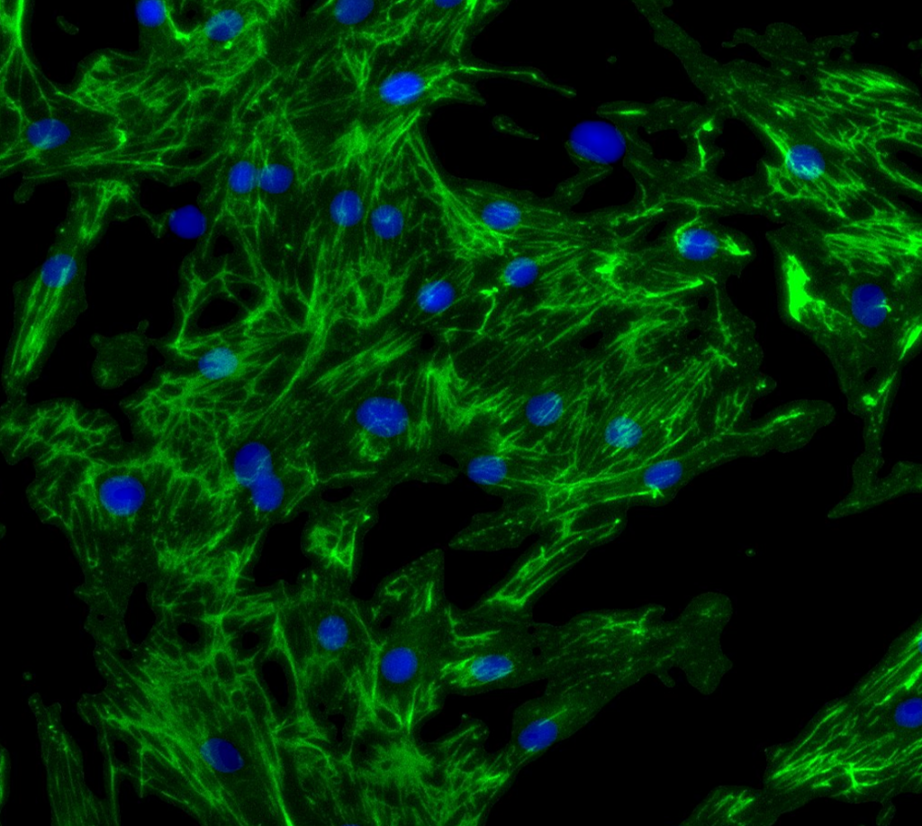
- Fibroblast migration - scratch assay; wound closure
- Epithelial migration - scratch assay; wound closure
- Fibrosis assays
Frequently Asked Questions – Fibrosis models
The right model depends on your target mechanism and organ of interest. For renal fibrosis, UUO and adenine diet models are the most widely used for evaluating antifibrotic efficacy, while STZ and IgA nephropathy models suit disease-specific mechanisms. For pulmonary fibrosis, bleomycin remains the gold-standard IPF model. Our team can help match your compound’s mechanism of action to the most translationally relevant model.
Reactive fibrosis (DOCA-salt + uninephrectomy) reflects diffuse interstitial fibrosis driven by chronic pressure/volume overload, while reparative fibrosis (isoprenaline-induced MI) reflects localized scar formation following acute cardiac injury. Testing across both models allows assessment of antifibrotic candidates at different stages of cardiac remodeling.
Yes. While renal fibrosis is our founding area of expertise, NEPHRIX Biosolutions has expanded to offer validated in vivo fibrosis models for the heart, lung, and liver, allowing sponsors to run multi-organ fibrosis programs with a single CRO partner.
Readouts are model-specific and typically include histological fibrosis scoring (e.g., Ashcroft score for lung, collagen content for heart), fibrotic marker expression (α-SMA, TGF-β1), and organ-specific functional parameters such as creatinine, BUN, or ALT/AST.
Yes. Our in vivo fibrosis models can be paired with in vitro fibrosis and migration assays (fibroblast and epithelial scratch assays) to provide mechanistic insight alongside whole-organism efficacy data, supporting a more complete translational package.
Yes, bleomycin-induced pulmonary fibrosis is the most widely validated and referenced model for idiopathic pulmonary fibrosis (IPF) research, reproducing key features of fibrotic lung remodeling used to assess antifibrotic efficacy.