Kidney fibrosis models built for translational precision
Renal fibrosis is where most antifibrotic candidates stop working
Fibrosis is the final common pathway of chronic kidney disease (CKD): progressive deposition of extracellular matrix replaces functional nephron tissue, driving an irreversible decline in renal function. It accounts for most of the progression toward end-stage renal disease, yet no antifibrotic therapy has reached full approval in CKD to date. The gap between preclinical efficacy and clinical translation remains the field’s central problem.
That gap is rarely about the biology alone, it’s just as often about model choice: an induction method that fails to reproduce the human injury pattern, a study window too short to capture true fibrotic remodeling, or endpoints that don’t align with what a clinical trial will actually measure. Model selection is the first translational decision in any antifibrotic program.
Model selection, at a glance
| Fast mechanistic screen | UUO |
|---|---|
| Systemic, function-linked CKD | Adenine diet |
| AKI-to-CKD transition | Unilateral IRI |
| Diabetic kidney disease | STZ |
| Glomerular disease, immune-driven CKD | IgA Nephropathy |
Which kidney fibrosis model fits your program?
A side-by-side view of NEPHRIX Biosolutions validated in vivo models: induction route, time to fibrosis, and the readouts each one supports.| Model | Species | Induction | Time to fibrosis | Key readouts | Best suited for |
|---|---|---|---|---|---|
| UUO - Unilateral Ureteral Obstruction | Mouse | Surgical ligation of one ureter | 7 - 14 days | Col1a1, α-SMA, hydroxyproline, F4/80 | Fast antifibrotic efficacy screening |
| Adenine Diet-Induced CKD | Mouse / Rat | Oral adenine-containing diet | 3 - 6 weeks | GFR, BUN/creatinine, tubulointerstitial fibrosis | Systemic CKD with functional decline |
| STZ-Streptozotocin-Induced DKD | Mouse / Rat | Single/multi-dose STZ injection | 8 - 16 weeks | Albuminuria, glomerulosclerosis, mesangial expansion | Diabetic kidney disease programs |
| Iga Nephropathy | Mouse | Immune-mediated glomerulonephritis induction | Variable, chronic | IgA deposition, proteinuria, glomerular injury | Glomerular disease, immune-driven CKD |
| IRI Ischemia-Reperfusion Injury | Mouse | Unilateral renal pedicle clamping (ischemia) followed by reperfusion | 4 - 8 weeks | KIM-1, NGAL, Col1a1, α-SMA, tubular injury score | AKI-to-CKD transition, nephroprotection |
Kidney Fibrosis in vitro assay, the DuoXProx™
Our proprietary renal co-culture system mimics epithelial-fibroblast crosstalk for more predictive in vitro fibrosis signal than monoculture assays.
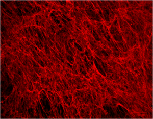
- Co-culture system: Reconstitution of the original cellular environment using Human primary cells
- Customizable stressors: Hormonal, Diabetic, Nephrotoxic or Hypoxic stressors availiable
- Multi-parameters analysis: Intracellular or secreted proteins quantification, gene expression, oxydative stress analysis.
- High-content imaging & biomarker analysis: Automated quantification of fibrosis markers on our CX7 CellInsight platform allowing consistent, blinded, image-based scoring.
Kidney fibrosis models — common questions
There isn’t a single “best” model, it depends on your question. UUO is the fastest and most reproducible for early antifibrotic screening. The adenine diet model better reflects systemic CKD with functional decline. Unilateral IRI is preferred when you need to study the AKI-to-CKD transition. We help you choose based on your compound’s mechanism and your program’s stage.
It’s model-dependent:
- UUO produces marked tubulointerstitial fibrosis within 7 – 14 days
- The adenine diet model typically requires 2 – 4 weeks
- Unilateral IRI takes longer, 4 to 8 weeks, to show progressive fibrosis and GFR decline, reflecting its slower, more clinically relevant disease course
UUO model is a surgical, obstruction-driven model producing fast, localized fibrosis in one kidney. UUO is ideal for rapid mechanistic screening. The adenine diet model is dietary and systemic, inducing bilateral kidney injury with measurable functional decline (GFR, creatinine), making it closer to progressive human CKD.
Core readouts include collagen deposition (hydroxyproline, Masson’s trichrome), fibrosis-related gene and protein expression (Col1a1, α-SMA), tubular injury markers (KIM-1, NGAL), and functional markers such as GFR, serum creatinine and BUN, combined with quantitative histopathology.
No, in vitro assays serve different purposes. In vitro assays like our DuoXProx™ co-culture system are well suited for early mechanism-of-action work and dose-response screening. In vivo models remain necessary to confirm efficacy in a physiological, systemic context before moving toward IND-enabling studies.
Why choose NEPHRIX Biosolutions?
Scientific Expertise
Ethical & 3R Considerations
Flexible Study Design
Gold standard and Specific Readouts
Why choose NEPHRIX Biosolutions?
Scientific Expertise
Ethical & 3R Considerations
Flexible Study Design
Gold standard and Specific Readouts